

Endometriosis: a primarily clinical diagnosis
Endometriosis can be diagnosed from puberty through to the menopause. However, there is often a delay in diagnosis (7 to 10 years between the appearance of the first symptoms and diagnosis).
The symptoms of endometriosis are generally more pronounced in women who menstruate monthly, and whose ovarian activity is not blocked by hormonal contraception. As a result, endometriosis is often diagnosed in teenagers, in young nulliparous women who stop using contraception to achieve pregnancy, or in multiparous women who opt for mechanical contraception (copper coil, tubal sterilization, etc.).
In other pauci- or asymptomatic women, the diagnosis is sometimes made by chance during an imaging scan or surgery performed for other reasons.
Clinical examination data
The diagnosis of endometriosis is primarily clinical. In most cases, an experienced examiner will be able to make the diagnosis long before any paraclinical examinations are carried out, thanks to a careful, targeted clinical examination and questioning.
Complementary examinations are designed to assess the severity of the disease (extension work-up), and to suggest appropriate treatment. They must be carried out by radiologists experienced in endometriosis. Their normality does not exclude the diagnosis, particularly in the case of superficial endometriosis.
Interrogation
It is fundamental and often sufficient to evoke the diagnosis. Its aim is to link the chronology of painful symptoms to the onset of menstruation. It should also look for the symptoms mentioned above (pelvic pain, dyspareunia, digestive and urinary symptoms, etc.).
Abdominal examination
It is often poorly performed, and generally causes non-specific pain (sensitive palpation of the colonic frame or pelvis). Nevertheless, when performed during a painful crisis, it can mimic a surgical abdomen. In addition, certain rare complications can genuinely lead to a surgical abdomen (occlusion, renal colic).
Speculum examination
It must be performed with care, as contact with the vaginal cul-de-sacs is generally painful. It is often normal, but may reveal a nodule of deep endometriosis infiltrating the vagina, in the form of a fibrous retraction, a budding lesion (Figure), or black microcysts.

Vaginal touch
It is essential, as it can confirm the diagnosis of deep endometriosis, but cannot be carried out on a virgin patient. Palpation may reveal several features:
- A soft but tender posterior vaginal cul de sac may point to superficial endometriosis of the cul de sac of Douglas.
- A fibrous nodule or budding lesion in the vaginal cul-de-sac, usually tender, may point to deep endometriosis.
- An enlarged, sensitive uterus may point to adenomyosis.
- A cystic mass may point to an ovarian endometrioma.
Rectal touch
It is particularly useful in cases of suspected deep retro-cervical endometriosis nodules, to assess the presence of infiltration of the rectum or parametrium.
Paraclinical examinations
Endovaginal pelvic ultrasound
Considered the first-line imaging test, ultrasound is most effective in the hands of an expert radiologist or gynecologist. It can identify :
- Ovarian endometriomas (finely granular ovarian cysts that persist over several cycles, unlike hemorrhagic cysts)
- Adenomyosis
- Deep endometriosis nodules of the uterosacral ligaments, bladder, rectum and distal sigmoid colon.
- Pelvic cystic collections such as obstructed and dilated fallopian tubes due to accumulation of menstrual blood (hematosalpinx), peritoneal pseudocysts due to adhesion formation, etc...
Abdominal ultrasound can be used to diagnose certain complications of endometriosis, such as ureteral stenosis with pyelocalic dilatation, kidney atrophy, extensive intra-abdominal endometriosis with ascites accumulation, etc.).
Magnetic resonance imaging (MRI)
Magnetic resonance magnetic resonance imaging (MRI) is a high-performance examination that enables not only diagnosis but also precise mapping of intra-pelvic and intra-abdominal endometriosis lesions. In our practice, it is virtually indispensable prior to any surgical treatment, and enables us to identify :
- Ovarian endometriomas containing blood have a very characteristic appearance: hypersignal in T1 sequence with fat saturation and hyposignal in T2 sequence (photo 1) ;
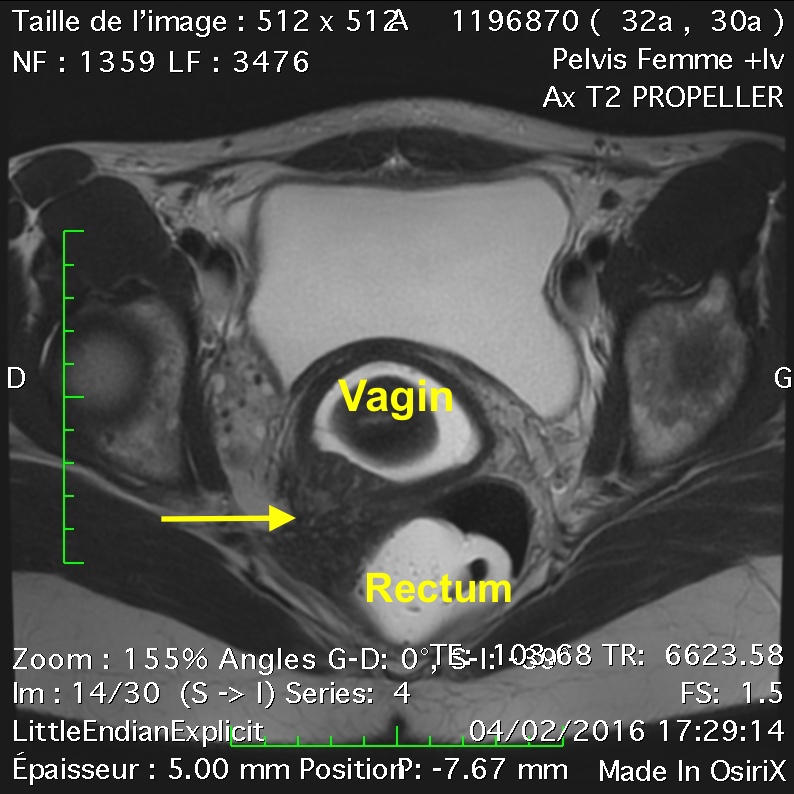
- Deep endometriosis nodules are star-shaped (hyposignal T1 and T2), retractile formations infiltrating neighboring organs (rectum, sigmoid colon, vagina, bladder, etc.) sometimes containing endometriosis microcysts (photo 2) ;
- Adenomyosis is confirmed by enlargement of the junctional zone or the presence of multiple hypersignal microspots in the myometrium (photo 3);
- Complications of endometriosis (hematosalpinx, ureteral stenosis with pyelocalic dilatation or hydronephrosis, infiltration of the parametrium or sacral roots, stenosis of the digestive tract, etc.).
Other examinations may be indicated in severe cases, depending on the location of endometriosis lesions visualized on ultrasound and MRI. They will be indicated on a case-by-case basis.
These include endorectal ultrasound and coloscanner in the case of colorectal endometriosis, cystoscopy, uroscanner or uroMRI in the case of urinary tract endometriosis, MRI and/or CT scan of the diaphragm in the case of diaphragmatic endometriosis.
No biological tests are specific. Increased CA125 levels may be observed in diffuse ovarian or peritoneal endometriosis, but this non-specific assay is not part of the work-up.
Research is currently underway to identify a blood marker that could indicate whether or not a woman has endometriosis, following a simple blood test.


Laparoscopy
If necessary, a diagnosis of certainty can be made by exploration of the abdominal cavity with targeted biopsy and anatomopathological analysis.
In cases of infertility, it enables prognostic classification according to AFSr (American Fertility Society revised), and sometimes treatment of superficial lesions.
Nevertheless, laparoscopy is not essential for initiating medical treatment in patients where endometriosis is strongly suspected after clinical examination and imaging studies. It should be reserved for situations of diagnostic doubt, and is therefore not recommended for the simple purpose of confirming imaging examinations when they are highly suggestive.
At present, laparoscopy is reserved mainly for the treatment of endometriosis lesions.
Saliva test
Endometriosis is often diagnosed when contraception is stopped
When stopping the contraceptive pill
Between the ages of 20 and 30, when women stop taking the contraceptive pill to try for pregnancy. Without medication, menstruation rapidly becomes very painful, and X-rays can reveal previously unrecognized endometriosis lesions.
After childbirth
or at any other time of life, when patients opt for a mechanical contraceptive method (copper coil, tubal ligation or tubal plugging); the resumption of menstruation without any drug braking unmasks previously unrecognized endometriosis lesions, which become symptomatic.
Get help from endometriosis specialists
For multidisciplinary endometriosis management, IFEM Endo supports you throughout your journey: medical and surgical treatment, medical follow-up, pain management.
Last updated on May 22, 2024 @ 11:23