Skip to content

Sacral plexus surgery
and sciatic nerve surgery

An endometriosis nodule on the sacral plexus or sciatic nerve is a relatively rare localization.

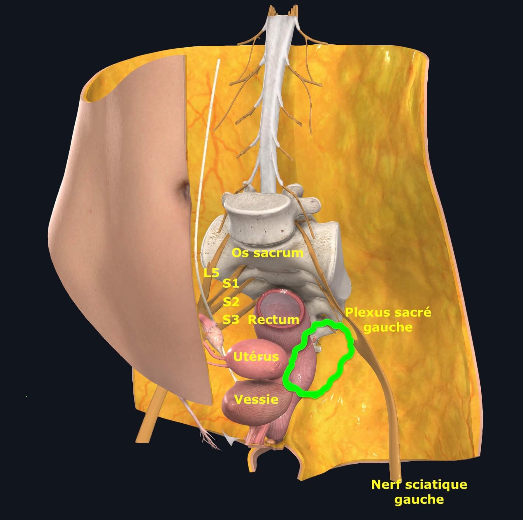
Deep endometriosis can affect the sacral plexuses, particularly when it spreads to the space lateral to the vagina and rectum, also known as the parametrium. The sacral plexus is located in the lateral, external and deep part of the parametrium, in contact with the pelvic wall. Endometriosis of the parametrium often infiltrates not only the sacral plexus, but also the vagina, rectum, ureters and even the bladder.

Endometriosis nodules of the sacral roots

Deep endometriosis nodules of the parametrium may compress, sheath or infiltrate large-diameter nerve structures such as the sacral roots, sciatic nerve, obturator nerve or pudendal nerve, as well as fine nerve structures such as the splanchnic nerves, hypogastric nerves, inferior and superior hypogastric plexuses.

Pelvic diagram with parameter and sacral plexus
Fig1: anatomical diagram of the female pelvis identifying nerve structures

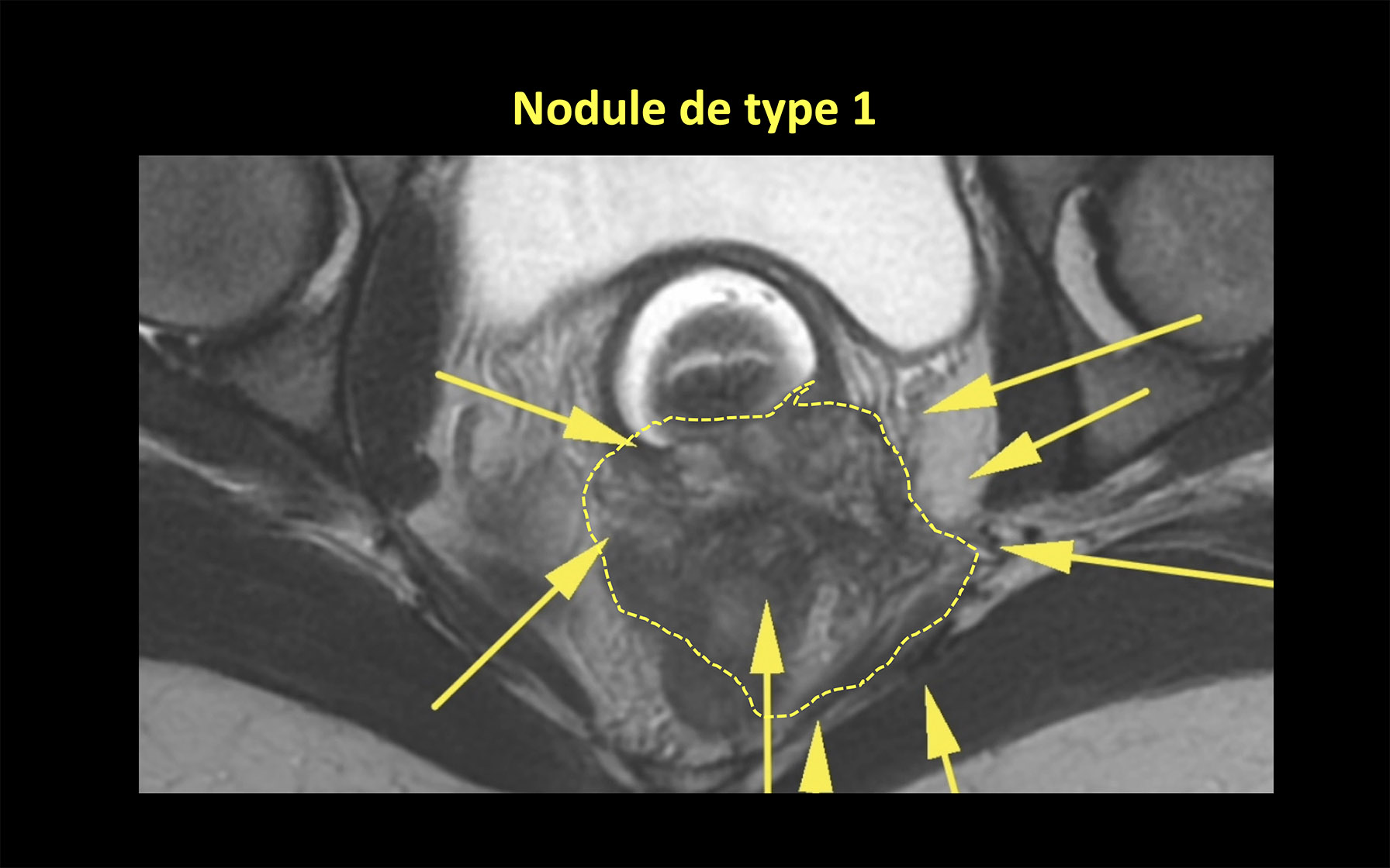
There are 2 types of parameter nodules:

  • Type 1. Large nodules occupying the medial part of the parametrium, compressing or infiltrating the sacral roots, but also the rectum and vagina. This is the most common type of sacral plexus involvement (85-90%). They cause pain in the buttock or perineum, associated with urinary or digestive disorders.
  • Type 2. More lateral nodules that compress or infiltrate the sciatic, pudendal and obturator nerves. These are rarer lesions (10-15%), manifesting as pain or motor disorders in the buttock and lower limb. Digestive and urinary disorders, on the other hand, are rarer.

Symptoms of sacral plexus endometriosis

Deep endometriosis nodules in the sacral plexuses may be responsible for several specific symptoms, particularly at the time of menstruation:

  • Sciatalgia: pain that starts in the buttock and runs down the back of the thigh to the bottom of the foot.
  • Pudendalgia: pain due to damage to the pudendal nerve, affecting the perineum from the labia majora to the clitoris and the lateral part of the anus.
  • Pain in the territory of the obturator nerve: pain on the medial side of the thigh, above the knee.
  • Bladder or rectal dysfunction: difficulty emptying the bladder, particularly during menstruation (patients are forced to push by contracting their abdominal muscles, or bend forward, to compress the bladder and help it empty) or slow transit and difficulty eliminating stools.
  • Vaginal dryness.

This type of clinical presentation of endometriosis is rare and often unrecognized by practitioners. Consequently, these symptoms are initially rarely attributed to endometriosis, but rather to rheumatological, osteoarticular or muscular causes.

Anatomy
of the
parameter and sacral plexus

The sacral plexus is made up of sensory and motor nerves. Some motor nerves are called "somatic" because they manage the voluntary motor response of skeletal muscle fibers in the lower limbs, buttocks or pelvis. Others are called "vegetative", as they are involved in the autonomic (involuntary) contraction of pelvic and abdominal organs (rectum, colon, bladder), erectile bodies, genital and skin glands, or vessels.

Somatic innervation (fig 4) of the pelvis comes from nerves or roots originating from the spinal cord, either from the lumbar area (denoted by L, from 1 to 5) or from the sacral area (S, from 1 to 4 or 5). The roots emerge from the spine through the vertebral holes and interconnect in branching structures, also known as "plexuses": lumbar and sacral.

The sacral plexus (fig. 5) is formed by the last lumbar root (L5) and the S1, S2, S3 and S4 or S5 roots. These nerve roots include afferent (information gathered in the periphery is directed to the spinal cord and brain) and efferent (information is sent to the muscles) fibers involved in sensory perception and voluntary movement of the lower limbs.

The sacral roots are located in the posterior pelvis, between the sacrum and the lateral pelvic wall, in front of the piriformis muscle (fig 6). They converge and give rise to larger-caliber nerves (4 mm to 1 cm):

  • The sciatic nerve (mainly with somatic fibers from L5, S1 and S2), which leaves the pelvis and travels to the posterior thigh, where it descends to the leg and foot. It is involved in motility of the leg and foot, and in sensitivity of the posterior aspect of the lower limb down to the foot.
  • The pudendal nerve (mainly S2, S3 and S4), which leads to the perineum (the area between the thighs containing the clitoris, vulva and anus) and to the external sphincter of the anus.
diagram of piriform nerve and sacral plexus nerves
Fig 6: Anatomical diagram showing the relationship between the piriformis muscle and the sacral plexus.

Finally, mainly vegetative fibers, originating from S2, S3 and S4, of small caliber (around 1-2 mm) interconnect to form a cobweb-like network, located deep in the parametrium, lateral to the rectum and below the ureter: the inferior hypogastric plexus. From this spider's web arise very fine nerves that reach the bladder, the floor of the vagina and the rectum: the splanchnic nerves. The splanchnic nerves control voluntary bladder emptying and the function of the internal sphincter of the urethra, but also influence rectal mobility and sexual functions.

The inferior hypogastric plexus also receives the hypogastric nerves, whose diameter is around 1-2 mm and which are involved, among other things, in the sensation of bladder fullness.

Adjacent to the sciatic nerve is the obturator nerve, which arises from the lumbar plexus (L2, L3 and L4), enabling movement of the thigh adductor muscles that move the thigh towards the midline of the body (bringing the thighs together), and providing sensitivity to the medial aspect of the thigh.

The anatomy of the pelvic nerves explains the localization of pain, motility disorders and pelvic organ function when the sacral plexus, sciatic, obturator and pudendal nerves are involved.

Assessment of sacral root endometriosis lesions

Surgery for endometriosis nodules of the sacral plexus cannot be considered without a careful preoperative workup.

MRI is the essential, mandatory reference examination, enabling the surgeon to visualize in 3 dimensions the location, dimensions, limits and volume of the endometriosis nodule. It determines whether neighbouring organs (rectum, ureter, bladder, vagina, piriformis muscle) have been affected, and enables surgery to be planned with the participation of a multidisciplinary team.

Urodynamic testing assesses bladder function, and can identify signs of nerve damage to the bladder. We reserve this examination for patients with obvious symptoms of altered bladder function, or where we suspect incomplete emptying of the bladder, justifying the implementation of a program of self-catheterization prior to surgery.

Anorectal manometry explores rectal function. In practice, we never perform it before surgery, and reserve it for patients with persistent defecatory problems after surgery.

Surgical treatment of sacral plexus endometriosis

Deep endometriosis lesions of the sacral plexuses and sciatic nerves are removed laparoscopically. The procedure is different for type 1 and type 2 nodules. Despite its complexity, we believe that this procedure is well standardized, and follows very precise steps, which ensure complete removal of the lesions, reduce the risk of intraoperative hemorrhage, and minimize the risk of postoperative functional sequelae.

To find out more about the surgical techniques used for the excision of endometriosis nodules of the sacral plexus and sciatic nerves, we suggest you consult the article published in 2020 by our team and available in open access:

The duration of surgery depends on the complexity of the deep-seated lesions, and in particular on the need to perform several complementary procedures on the rectum, ureter, vagina or bladder. Surgery for endometriosis of the sacral plexuses involves several highly complex procedures in a single operation.

The duration of such surgery can vary from 2 to 8 hours.

It's important to understand that it's sometimes impossible to preserve certain nerves during the removal of large endometriosis nodules from the parametrium, especially the finer ones. Sacrificing these small nerves can lead to at least temporary impairment of bladder and rectal function. 

In some cases, however, when endometriosis affects both parameters, we are obliged to perform a complete resection on the side where the disease appears most aggressive, and to perform the minimal procedure on the side least affected, in order to avoid complete denervation of the bladder, rectum or vagina. While small-calibre pelvic nerves may be sacrificed unilaterally, surgery on large-calibre nerves such as the sciatic or obturator nerve must be as conservative as possible to avoid the development of major motor or sensory disorders.

Hospitalization

The length of hospital stay varies from 3 to 7 days, depending on the type of surgery performed and the post-operative course. Recovery is faster if the surgery does not involve suturing the rectum or ureters.

Postoperative follow-up of sacral plexus endometriosis

The post-operative course can be marked by several immediate complications, which must be clearly presented before surgery:

Bladder atony

Voluntary emptying of the bladder is achieved by a command transmitted to the bladder via the splanchnic nerves. The splanchnic nerves are bilateral, but their participation (dominance) may be unbalanced. They can be affected both by the endometriosis nodule (infiltration, compression, irritation, in which case symptoms precede surgery) and by the surgical procedure. During surgery, the very fragile splanchnic nerves may be removed en bloc with the nodule, or sectioned. 

Even when preserved, splanchnic nerves can be affected by heat diffusion from the energies used to coagulate vessels, or stretched during dissection, leading to neuropraxia. The term neuropraxia is used to describe moderate nerve damage resulting in temporary impairment of nerve function, and corresponds to a temporary loss of the nerve's myelin sheath, without any associated axonal damage. This demyelination slows the speed of conduction of the electrical impulse within the nerve, and consequently impairs the transmission of information through the nerve. 

The prognosis is favorable, with complete recovery usually within a few weeks or months, due to reformation of the myelin sheath. The clinical result is difficulty in emptying the bladder, which permanently retains a variable quantity of urine. If the volume of urine remaining in the bladder at the end of a micturition (post-micturition residue or PMR) exceeds 100ml, patients must use small bladder catheters to empty the bladder several times a day (usually 5 or 6 times), at set times, and after attempting voluntary micturition. Bladder function improves progressively, over 4-6 weeks post-operatively, and self-catheterization can be discontinued when RPM consistently falls below 100ml. 

When self-catheterization is indicated, it is taught before discharge from the clinic. Patients are asked to regularly record the volumes evacuated by catheterization (voiding calendar) and to communicate these to the surgeon.

The incidence of bladder atony requiring systematic self-catheterization is around 25% at discharge, decreasing to around 5% one year after surgery. Recovery of bladder function is explained by the regression of neuropraxia, edema and local inflammation due to surgery.

Neuropathic pain, paresthesia or hyperesthesia in sciatic, obturator and pudendal nerve territories.

These pains are the clinical expression of irritation, nerve edema or somatic nerve neuropraxia, and manifest as electrical discharges, tingling, or painful sensations triggered by minimal stimulation (contact with clothing). 

These symptoms require specific treatment with drugs that interfere with nerve transmission, such as pregabalin, amitriptyline and gabapentin, administered over several weeks or months. 

These symptoms are present in around 17% of cases one year after surgery.

Long-term data are encouraging, both in terms of clinical improvement and fertility. It is important to note that women with deep endometriosis involving the sacral plexus or sciatic nerve more rarely have associated ovarian or tubal damage. This could explain the very high pregnancy (77%) and postoperative birth rates, with spontaneous conceptions in half the cases. 

This information should encourage patients to undergo surgery, even if they are trying to become pregnant. Indeed, scientific data do not support the use of in vitro fertilization in this situation, which also delays surgery and makes it technically more complex due to the continued growth of lesions.

Endometriosis
of the sacral plexuses
in figures

20-25

patients with sacral plexus damage operated on every year

82,7%

of patients with sacral plexus involvement also have a severe rectal lesion

47%

of patients with sacral plexus involvement also have a urinary tract lesion

Bibliography

Roman, Merlot et al. JMIG 2020 

Get help from a sacral plexus endometriosis specialist

IFEM Endo, a center specializing in the management of endometriosis and complex forms of the disease, will accompany you on your journey of care for sacral plexus endometriosis.

Last updated on May 1, 2021 @ 08:55

Post-operative emergency

After your surgery, if you have any concerns after you return home and before your post-operative visit, you can contact us quickly using the form below.

Please use this emergency form only in the immediate post-operative period.


Your doctor? *
Date of your intervention? *

Please explain your problem below:

*Mandatory fields.

Post-operative follow-up

After your surgery and post-operative visit, if you encounter a problem with your treatment or have a question or query, you can contact us quickly using the form below.


Your doctor? *
Date of your intervention? *

Please explain your problem below:

*Mandatory fields.

Make an appointment: patient follow-up

If you have already consulted an IFEM Endo doctor and would like to make a new appointment for your follow-up, you can contact us quickly using the form below.


Your doctor? *
Please explain your problem:

*Mandatory fields.

Contact: Research team

If you wish to contact the research team, please complete this form.

*Mandatory fields.